Most people never think about their retina until something goes wrong. Vision simply feels automatic. You wake up, open your eyes, recognise faces, read messages, drive through London traffic, notice colours, movement, depth, and light, all without consciously thinking about the complex biological system making it possible.
But behind every sharp image sits one of the most important structures in the human eye: the retina. Often described as the “film” or “sensor” of the eye, the retina is responsible for converting light into electrical signals that the brain can understand. Without it, vision itself would not exist. Retinal health plays an important role in everything from routine eye assessments to advanced vision correction planning. Understanding how the retina works can help patients recognise why regular eye examinations are so important, particularly as we age.
What Is the Retina
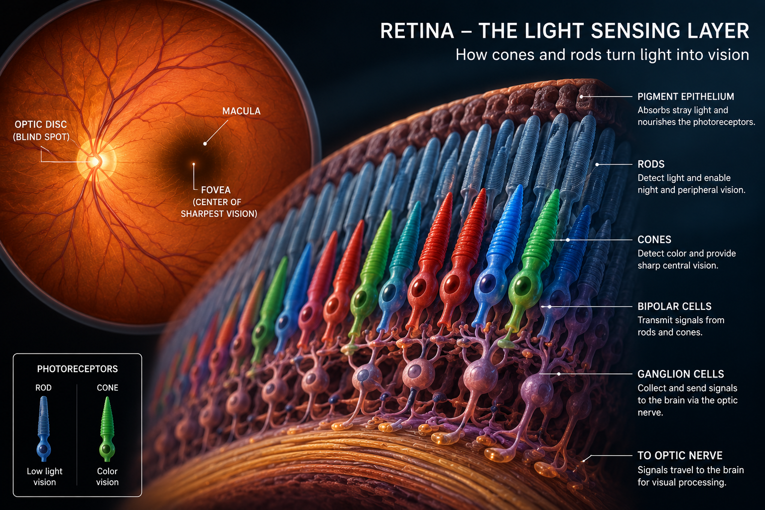
The retina is a thin, light-sensitive layer lining the back of the eye.
Its job is to capture incoming light and transform it into electrical signals that travel through the optic nerve to the brain. Only once the brain processes these signals do we actually “see” the world around us. The retina contains highly specialised cells called rods and cones.
These cells allow us to detect colour, contrast, movement, brightness, and detail under different lighting conditions. At the centre of the retina sits the macula, the area responsible for our sharpest central vision. Within the macula lies the fovea, the point where visual detail becomes most precise. Without the retina functioning correctly, even perfectly healthy glasses, lenses, or corneas cannot produce clear vision.
How the Retina Actually Works
The retina works more like a living neurological processor than a simple camera sensor. Every second, light enters the eye through the cornea and lens before landing precisely on the retinal surface. The retina then begins an extraordinary chain reaction.
Step One – Detecting Light
Specialised photoreceptor cells absorb incoming light. Cones allow us to see fine detail and colour during daylight conditions, while rods help us see in dim environments and at night. This is why colours become muted in darkness, rods are active, but cones require brighter light to function properly.
Step Two – Processing Information
The retina does not simply capture images. It actively processes them. Different retinal layers organise contrast, brightness, movement, and spatial information before passing signals deeper into the visual system.
Step Three – Sending Signals to the Brain
Once processed, electrical signals travel through the optic nerve directly into the brain. The brain then reconstructs these signals into the images we experience as vision. In many ways, the retina acts as the bridge between the physical world and human perception itself.
Why Sharp Vision Depends on the Retina
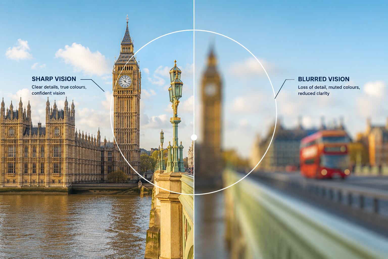
For clear vision to occur, light must land precisely on the retinal surface. In short-sightedness, long-sightedness, and astigmatism, the eye’s focusing system causes light to land either in front of or behind the retina instead of directly on it. That is why vision becomes blurred.
Glasses, contact lenses, laser eye surgery, and lens replacement procedures all work by changing how light focuses so that images land correctly on the retina once again. The retina itself remains central to all forms of visual correction.
The Structure of the Retina
The retina consists of multiple microscopic layers working together simultaneously. Although highly complex scientifically, its structure can be understood through three main functional areas.
Protection and Support
The outer retinal layer helps absorb excess light and supplies oxygen and nutrients to the retinal cells. This support system prevents visual distortion and keeps retinal tissue functioning properly.
Light Detection
This area contains the rods and cones responsible for capturing visual information. The macula contains an especially dense concentration of cones, allowing detailed reading, facial recognition, and fine central vision.
Signal Transmission
Once light is converted into electrical information, retinal nerve cells organise and transmit signals into the optic nerve, which carries information directly to the brain. Interestingly, the optic nerve creates a natural blind spot where it exits the eye because no photoreceptor cells exist in that exact location.
What Happens When the Retina Becomes Damaged
Because the retina contains delicate nerve tissue, damage can significantly affect vision. Some retinal conditions develop gradually over years, while others appear suddenly and require emergency treatment.
Common warning signs include:
- Distorted or blurred vision
- Dark patches in central vision
- Flashes of light
- Sudden floaters
- Loss of peripheral vision
- Reduced colour perception
- A dark “curtain” appearing across vision
Sudden retinal symptoms should never be ignored. Flashes, floaters, or shadow-like vision loss can sometimes indicate retinal detachment, which requires urgent ophthalmic treatment.
Common Retinal Conditions
Age Related Macular Degeneration
Macular degeneration affects the central retina and becomes increasingly common with age. Patients often notice distorted lines, difficulty reading, blurred central vision, or faded colours.
Diabetic Retinopathy
High blood sugar levels can damage the tiny blood vessels supplying the retina. Over time, this may cause bleeding, swelling, blurred vision, or progressive visual loss.
Retinal Detachment
This occurs when the retina separates from the underlying tissue supporting it. It is considered a medical emergency because untreated detachment can lead to permanent sight loss.
Macular Oedema
Fluid accumulates within the central retina, causing blurred or distorted central vision.
Retinitis Pigmentosa
A rare inherited condition affecting the retinal photoreceptor cells, often beginning with night blindness and progressive tunnel vision.
Can the Retina Repair Itself
The retina has limited natural regenerative ability compared with many other tissues in the body. However, modern ophthalmology has developed several advanced treatments that can preserve vision or slow further damage.
Depending on the condition, treatment options may include:
- Laser retinal therapy
- Retinal surgery
- Anti-VEGF injections
- Vitreoretinal procedures
- Electronic retinal implants
- Emerging stem cell therapies
Early diagnosis remains one of the most important factors influencing visual outcome. Many retinal diseases respond significantly better when identified before major vision loss occurs.
The Retina and Modern Vision Correction
Retinal health is also an important part of assessing suitability for laser eye surgery and lens procedures.
Before treatments such as SMILE Pro, Femto-LASIK, PRESBYOND, ICL surgery, or refractive lens exchange are considered, detailed retinal examinations help ensure the eye is healthy enough for surgery.
At EuroEyes London, advanced diagnostic imaging allows surgeons to examine both the front and back of the eye in exceptional detail before recommending treatment. That level of analysis is important because excellent visual outcomes depend on far more than prescription strength alone.
Why Regular Eye Examinations Matter
One of the challenges with retinal disease is that many conditions begin silently. Patients may notice no symptoms until vision has already changed significantly. Routine eye examinations therefore play an important role not only in updating prescriptions, but also in detecting early retinal disease before symptoms become severe.
For many people, retinal imaging can reveal wider health issues such as diabetes, vascular disease, or high blood pressure long before they would otherwise be diagnosed. The retina is not simply part of the eye. In many ways, it is one of the only places in the human body where delicate nerve tissue and blood vessels can be observed directly in real time. And that makes it one of the most important windows into overall visual health.