Keratoconus is a condition that raises immediate questions for anyone diagnosed with it. The most common one is simple but important: is there a cure. People naturally want to know whether their vision can return to normal, whether the condition will stop progressing and whether treatment can reverse the changes already taking place in the eye. This guide explains what modern medicine can achieve, how keratoconus is managed and what patients can realistically expect from long-term care.
What keratoconus actually is
Keratoconus affects the shape and structure of the cornea. Instead of remaining smooth and rounded, the cornea becomes thinner and gradually bulges forward. This distorts vision and can lead to irregular astigmatism, ghosting, glare and a noticeable decline in clarity. The condition often begins in the teenage years or early adulthood and can progress slowly or unpredictably.
Although keratoconus can sound alarming, it is a condition that modern eye care understands well. Early diagnosis and the correct treatment plan can slow, stabilise or control its progression and in many cases protect vision for the long term.
Can keratoconus be cured
There is currently no treatment that reverses keratoconus completely or restores the cornea to its original pre-condition structure. In this sense, keratoconus cannot be cured in the traditional meaning of the word. However, many patients misunderstand what this actually means. The absence of a cure does not mean the absence of effective treatment.
Modern techniques are highly successful at stopping or slowing progression, improving visual quality and protecting the eye against further change. In practice, this means that many patients live full, visually stable lives with the help of early intervention and ongoing care.
The modern goal is not cure. It is stability, clarity and long-term preservation of vision.
What modern treatment can achieve today
Keratoconus is managed through a combination of treatments. Each method has a purpose and plays a role in strengthening, reshaping or supporting the cornea.
Corneal cross-linking (CXL)
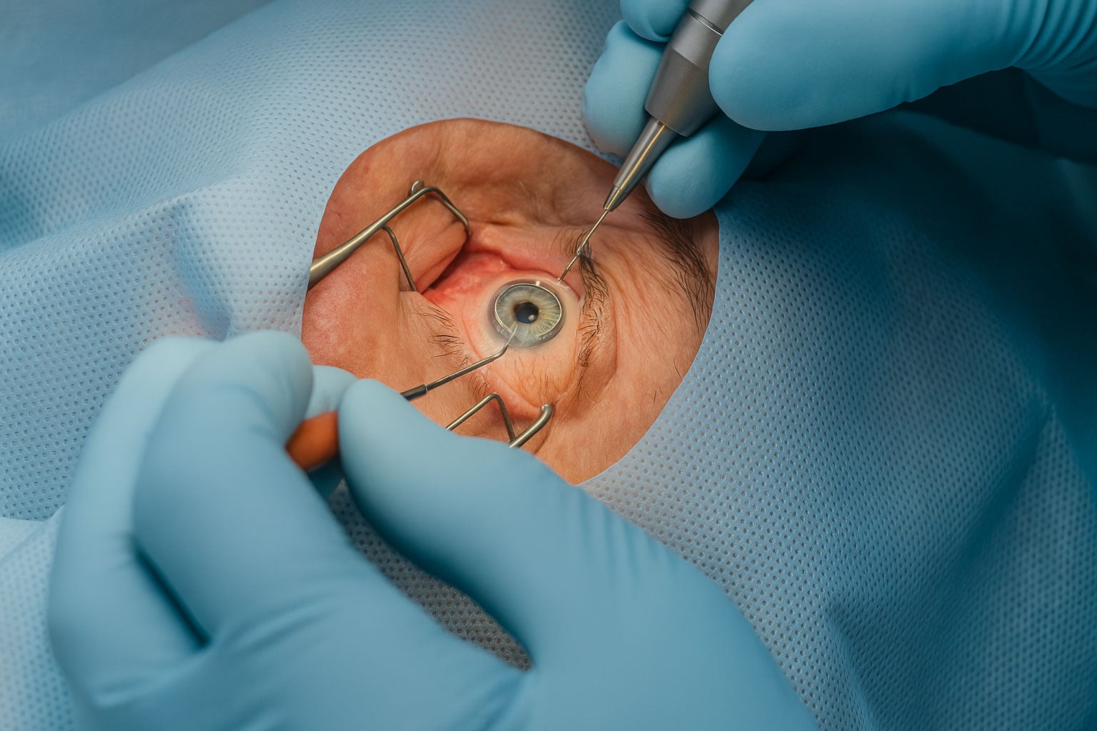
CXL is the primary treatment used to stop keratoconus from getting worse. The technique strengthens the corneal tissue using riboflavin drops and controlled ultraviolet light. This process creates new collagen bonds inside the cornea, helping it resist further thinning or bulging.
CXL does not reverse existing changes but it is highly effective at halting progression. Many patients experience a lifetime of stability after treatment. Early detection is important, because CXL works best before the cornea becomes severely misshapen.

PRK for mild keratoconus or post-CXL refinement
In selected cases, superficial laser treatment can be used to improve the corneal surface once keratoconus has been stabilised by CXL. This can help reduce irregularities and improve visual clarity. It is not suitable for everyone, but it plays an important role for certain patients who want to reduce dependence on specialty contact lenses.
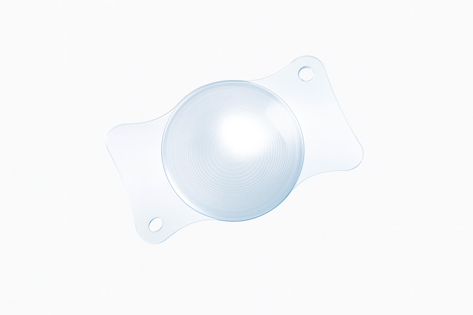
Implantable contact lenses (ICL)
ICLs are a powerful option for patients who cannot achieve clear vision with glasses or standard lenses. An ICL is placed inside the eye to correct short sight, long sight or astigmatism. For keratoconus patients who have stable corneas but persistent refractive error, ICLs can offer sharp, comfortable vision without reshaping the cornea.
Specialty contact lenses
Keratoconus-specific lenses, including scleral and hybrid lenses, can restore excellent visual quality in many cases. These are often used while waiting for CXL or alongside long-term management.
Corneal grafts
In advanced cases where the cornea becomes too thin or distorted to support other treatments, a corneal transplant may be considered. This is far less common today due to early diagnosis and the success of CXL, but it remains an option for complex cases.
KeraNatural and KeraRings: Additional Options for Keratoconus Management
While cross-linking, PRK, ICLs and scleral lenses form the foundation of keratoconus care, some patients benefit from additional techniques designed to reshape or support the cornea. Two such options are KeraNatural and KeraRings. These treatments are not cures, but they can play an important role in improving corneal stability, vision and quality of life when used in the right circumstances.
KeraNatural (Topography-Guided Laser Refinement After CXL)
KeraNatural refers to a customised surface-laser optimisation performed after corneal cross-linking once the cornea has stabilised. The goal is not to treat keratoconus directly, but to improve how the corneal surface bends light.
It is particularly useful for patients with stable keratoconus who still experience irregular astigmatism, those who cannot obtain good vision with glasses, and people who want to reduce dependence on specialty contact lenses.
A topography-guided excimer laser gently smooths irregular high points on the cornea, reducing visual distortion without compromising corneal strength. KeraNatural is always performed after CXL, not before.
Benefits include reduced ghosting, glare and irregular astigmatism, the potential for better vision with or without lenses and improved outcomes if implantable lenses are used later. However, it is not suitable for advanced thinning, results can vary depending on the corneal shape and it requires a stable post-CXL cornea.
KeraRings (Intracorneal Ring Segments)
KeraRings are small, curved implants made of biocompatible PMMA material. They are placed within the cornea to reshape it from the inside. Unlike a graft or laser procedure, KeraRings do not remove any tissue – they simply alter the biomechanics of the cornea.
They are considered for patients with moderate keratoconus who struggle with glasses or normal contact lenses, or where the cornea is too irregular for PRK but not thin enough to require a transplant.
Using a femtosecond laser, channels are created within the cornea and one or two semicircular ring segments are inserted to flatten and regularise the corneal shape. This can reduce cone steepening, improve symmetry and enhance the effectiveness of glasses or contact lenses.
KeraRings can improve corneal shape and visual quality, are reversible and may delay or avoid the need for a corneal transplant. They do not, however, stop keratoconus progression – cross-linking is still required and results depend on accurate ring selection. They are not suitable for severely thin or scarred corneas.
Where KeraNatural and KeraRings Fit into the Treatment Pathway
A common modern sequence looks like this:
- CXL to stop progression
- KeraRings, where appropriate, to reshape the cornea
- KeraNatural (topography-guided PRK) to refine the surface
- Implantable contact lenses (ICLs) for final visual correction if required
Not every patient requires all of these steps, but having access to them allows the surgeon to tailor care precisely to the eye’s structure and long-term needs.
Why timing matters
Keratoconus is a condition where time plays a significant role. Early diagnosis allows for early cross-linking, which can prevent further thinning of the cornea. Once the cornea stabilises, additional treatments such as PRK or ICLs become safer and more effective. Patients who delay diagnosis sometimes experience faster progression during their younger years, when the cornea is more prone to change.
This is why any suspicion of keratoconus should be assessed promptly, particularly in teenagers and young adults.
How keratoconus progresses
Progression varies from person to person. Some patients notice gradual changes over several years, while others experience faster shifts in prescription or shape. Regular corneal topography scans are essential, as they allow the clinic to track subtle changes long before they are visible in day-to-day life.
EuroEyes uses advanced imaging tools to measure corneal thickness, curvature, biomechanics and stability. These measurements allow for early intervention, which can make a significant difference to long-term vision.
How EuroEyes assesses keratoconus
The diagnostic part of the journey is one of the most important. EuroEyes carries out a detailed scan of the cornea using high-resolution imaging technologies that map the shape, thickness and biomechanical behaviour of the cornea. This helps identify keratoconus at its earliest stages, sometimes even before a patient notices changes in their vision.
Once the scans are complete, the surgeon will explain the degree of progression, discuss the likelihood of further change and outline the most suitable treatment pathway.
Treatment pathways that work
Keratoconus care is personalised. A typical pathway may look like this:
- Early-stage progressive keratoconus: CXL to prevent further change
- Stable post-CXL cornea with irregular vision: PRK in selected cases
- Residual short sight or astigmatism: ICLs for sharper vision
- Visual distortion or lens intolerance: specialty scleral lenses
- Advanced thinning: consideration of a partial or full corneal graft
Most patients do not need a transplant. With early detection and timely CXL, the majority achieve stable long-term outcomes.
| Treatment | Main Purpose | When It Is Used | Benefits | Limitations |
|---|---|---|---|---|
| Corneal Cross-Linking (CXL) | Stops progression | Early to moderate keratoconus with signs of change | Strengthens the cornea and helps prevent further thinning | Does not reverse existing distortion |
| PRK (Surface Laser) | Improves surface regularity | After CXL once the cornea is stable | Can smooth irregularities and improve visual quality | Not suitable for advanced thinning; not a stand-alone keratoconus treatment |
| Implantable Contact Lenses (ICLs) | Corrects vision without reshaping the cornea | Stable keratoconus with significant refractive error | Provides sharp, predictable vision with a reversible implant | Does not treat the underlying corneal structure |
| Scleral or Hybrid Lenses | Provides clear vision over an irregular cornea | Mild to advanced keratoconus when glasses are ineffective | Excellent clarity and comfort for many patients | Requires specialised fitting and regular lens care |
| Corneal Transplant | Replaces damaged or severely thinned corneal tissue | Advanced keratoconus with scarring or extreme thinning | Can restore corneal shape and improve vision | Involves surgery with longer recovery and ongoing graft management |
Long-term expectations
Living with keratoconus today is very different to a decade ago. With modern diagnostic tools and treatments, most patients maintain stable vision and avoid progression to the advanced stages. Regular monitoring remains important, especially for younger patients or those with a family history of the condition.
Many patients find that a combination of CXL and appropriate refractive correction allows them to continue driving, working, studying and living without significant restrictions.
When keratoconus becomes serious
Keratoconus becomes more serious when the cornea thins to the point of structural risk or when vision can no longer be corrected with glasses or lenses. This is far less likely to occur when the condition is detected and treated early. The important message is that keratoconus is manageable, but it requires attention and regular review.
What happens if keratoconus is left untreated
If keratoconus progresses without treatment, the cornea may continue to thin and bulge. Vision can become increasingly distorted and, in some cases, a corneal transplant may eventually be required. This progression is much less common today due to the availability of CXL, but it reinforces why early diagnosis is important.
Frequently Asked Questions
Can keratoconus be cured
Keratoconus cannot be reversed, but it can be effectively stabilised and managed. Modern treatments such as corneal cross-linking prevent further progression in most patients, and additional options like implantable lenses or specialist contact lenses can improve vision significantly.
Does keratoconus always get worse
Keratoconus often progresses during the teenage years and early adulthood, but the rate varies from person to person. Cross-linking can halt progression, which is why early diagnosis is important. Many patients remain stable once treated.
What is the best treatment for keratoconus
The best treatment depends on the stage of the condition. Cross-linking is used to stop progression, while PRK can improve the corneal surface in suitable cases. Implantable contact lenses are chosen to correct vision once the cornea has stabilised. Advanced cases may require specialist scleral lenses or, in rare instances, a corneal graft.
Can glasses or contact lenses fix keratoconus
Glasses can help in the early stages, but they often struggle to correct irregular astigmatism. Specialist lenses such as scleral or hybrid lenses can offer excellent vision even when the cornea is more distorted.
Is keratoconus hereditary
There is a genetic component, but not everyone with a family history develops the condition. It is more common in people who rub their eyes frequently or who have conditions that affect collagen stability. Regular screening is recommended for close relatives.
Can I get laser eye surgery if I have keratoconus
Conventional laser vision correction is not suitable for corneas affected by keratoconus. However, PRK may be considered after cross-linking in selected cases to improve surface regularity. For visual correction, implantable lenses offer a safe alternative.
Does keratoconus lead to blindness
Keratoconus does not cause total blindness, but it can lead to significant visual impairment if left untreated. With modern treatment such as cross-linking, most patients retain useful, stable vision throughout their lives.
How quickly does keratoconus progress
Progression can occur slowly over years or more rapidly in younger patients. Regular topography scans help detect subtle changes before they affect daily vision. Early treatment provides the best long-term outcomes.
When should I consider cross-linking
Cross-linking is recommended as soon as progression is confirmed. Treating early helps protect the cornea before the shape becomes excessively distorted. Many young patients undergo CXL shortly after diagnosis.
Will I need a corneal transplant
Most patients do not require a transplant. With early detection and timely cross-linking, the risk of reaching the advanced stages that require surgery has reduced significantly over the past decade.
A final word from your EuroEyes surgeon
If you have recently been diagnosed with keratoconus or are concerned about changes in your vision, I encourage you to arrange a detailed assessment. Understanding the stage of the condition is the first step towards stabilising it. With early intervention and a clear treatment plan, keratoconus can be managed very effectively. My team and I are here to support you at every stage of your journey and to help protect your vision for the long term.